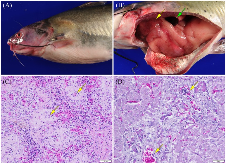
Figure 5.
Photographs of channel catfish infected by vAh showing (A) external surfaces that are exhibiting congestion/hemorrhage around the head/pectoral fin and within the eye and (B) the celomic cavity that has internal organs moderately congested and enlarged, a congested/hemorrhagic spleen (green arrow), and multifocal pale foci corresponding to areas of necrosis (yellow arrow) scattered over the liver (photographs courtesy of Dr. Wes Baumgartner, Mississippi State University) as well as photomicrographs of a channel catfish infected by vAh strain ML09-119 showing (C) a section of spleen with splenic ellipsoids (arrows) that are edematous and ellipsoidal arteries that are lined by degenerating as well as necrotic endothelial cells and (D) a section of liver with edema and necrosis of pancreatic acinar tissue surrounding branches of the hepatic portal vein (arrows).

