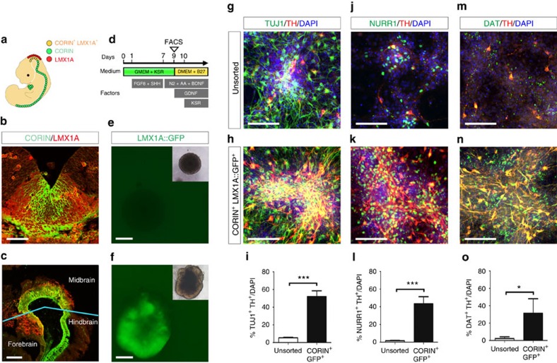
Figure 1. Purification of mDA progenitors by co-expression of CORIN and LMX1A::GFP.
(a) Schematic diagram of CORIN and LMX1A expression during early development of mouse. (b,c) Immunohistochemical images for CORIN (green) and LMX1A (red) in coronal and sagittal sections of E11.5 fetal mouse. Scale bars, 50 μm (b) and 200 μm (c). (d) Schematic diagram of neuronal differentiation from mESCs. (e,f) LMX1A::GFP expression of the serum-free floating culture of embryoid body-like aggregates with the quick reaggregation (SFEBq)-cultured mESC aggregates on day 2 (e) and day 9 (f). Scale bars, 200 μm. Insets indicate bright-field images of the aggregates. (g–o) Immunofluorescence images of the cells from unsorted and CORIN+LMX1A::GFP+ cells for TUJ1 (green), NURR1 (green), DAT (green), TH (red) and 4′, 6′-diamidino-2-phenylindole (DAPI; blue) on day 14. Scale bars, 70 μm. Quantification of TUJ1+TH+, NURR1+TH+ and DAT+TH+ cells in unsorted cells (n=4) versus CORIN+LMX1A::GFP+ cells (n=4) on day 14. Asterisks indicate statistical significance as determined by Student's t-test, *P<0.05 and ***P<0.001. Error bars indicate s.e.m.

