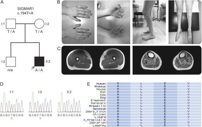
Figure. Family segregation, conservation of the SIGMAR1 variant, and clinical images of the proband.
(A) Segregation of the SIGMAR1 variant c.194T>A in the family; genotypes are indicated below tested individuals. (B) Photographs of the proband show atrophy of intrinsic hand muscles, clawed hands, flexed knee posture (characteristic of knee bobbing), and atrophy of leg and foot muscles. (C) Axial T1-weighted MRI of the mid-thighs (left) and mid-legs (right) demonstrate normal appearance of the thigh muscles and atrophy of all lower leg muscles with mild fatty replacement especially of tibialis anterior, tibialis posterior, soleus, and peroneal muscles. (D) Sanger sequencing electropherograms demonstrate sequence variants in the proband and his parents. (E) Conservation of leucine (L) at amino acid position 65 of the σ-1 receptor encoded by SIGMAR1; a subset of 16 species were chosen, representing the 100 species available at the USCS browser.

