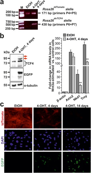
Figure 2.

The Rosa26dnTCF4 allele suppresses Wnt signaling in MEFs. (a) PCR genotyping of MEFs isolated from Rosa26CreERT2/tdTomato embryos treated with solvent [ethanol (EtOH)] or 4‐OHT for 4 days to induce Cre‐mediated recombination of the transcription blocker. PCR of the Rosa26tdTomato and Rosa26dnTCF4 allele produced 171 bp or 430 bp DNA fragments, respectively. M, molecular weight markers. Notice that since CreERT2 is expressed from the Rosa26 locus, EGFP‐dnTCF4 is produced from a single Rosa26 allele. (b) Immunoblotting (left) and qRT‐PCR analysis (right) of MEFs isolated from Rosa26CreERT2/tdTomato embryo treated with EtOH or 4‐OHT for 4 days. EGFP‐dnTCF4 fusion protein (displaying lower mobility than endogenous Tcf4) was detected using anti‐TCF4 (red arrows) and anti‐EGFP antibodies. Notice that Tcf4 migrates in denaturing conditions as a double band. Immunoblotting with an anti‐α‐tubulin antibody was used as a loading control. Diagram shows results of the qRT‐PCR analysis of Wnt‐responsive genes Axin2, Nkd1, and Troy in MEFs stimulated overnight with recombinant Wnt3a. The “housekeeping” gene β‐actin (Actb) was also included in the test. The quantities of input cDNAs were normalized to Ubiquitin B (Ubb). The expression level of a given gene in unstimulated cells was arbitrarily set to 1. Error bars represent standard deviations (SDs); **P < 0.01 (Student's t‐test). (c) Fluorescent microscopy images of MEFs derived from Rosa26CreERT2/tdTomato embryo treated with EtOH (left panel) or 4‐OHT for 4 and 14 days. Nuclear EGFP‐dnTCF4 fusion protein was detected by immunocytochemical staining using an anti‐GFP antibody (green fluorescence); tdTomato protein was visualized by its native red fluorescence. Specimens were counterstained using the DAPI nuclear stain (blue). Original magnification: ×400.
