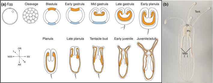
Figure 2.

Development and morphology of Nematostella. (a) Schematic of Nematostella developmental stages. Modified from Ormestad et al. Ref 90. Orange represents presumptive endoderm in blastula and the endoderm in all subsequent stages. The outer ectoderm is white, and the aboral ectodermal domain is in light blue. (b) DIC image taken by Aldine Amiel of juvenile polyp. The mouth (oral opening) is indicated by (Or), the tentacles by (tent), the pharynx by (Ph), and the mesenteries by (mes). Oral is up in all images.
