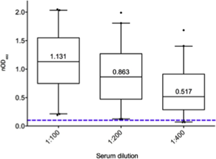
Figure 2.

BKPyV IgG serology of 42 healthy individual participants. Normalized BKPyV IgG antibody levels are shown at 1:100, 1:200 and 1:400 dilutions (median, box shows 25th, 75th percentiles; whiskers 5% and 95%). Positive serological status was defined as OD492nm ≥0.100 (dotted line) at the 1:200 dilution. BKPyV, BK polyomavirus; nOD, net optical density.
