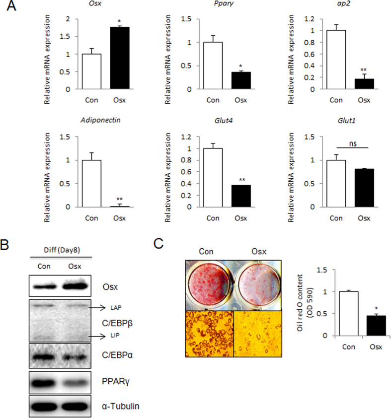
Figure 2. Overexpression of Osterix inhibits adipogenesis in 3T3-L1 cells.
3T3-L1 cells were transfected with empty vector (Con) or Myc-Osterix expressing plasmid (Osx). Cells were then cultured in adipogenic medium for 8 days. (A) The mRNA expression of Osterix, Pparγ, ap2, Adiponectin, Glut4, and Glut1 was determined by real-time PCR and normalized to Gapdh. (B) The protein expression of Osterix, C/EBPβ, C/EBPα, and PPARγ was confirmed by immunoblotting. α-tubulin was used as a loading control. For C/EBPβ, liver-activating protein (LAP) and liver-inhibitory protein (LIP) are indicated. (C) Oil red O staining of lipid droplets in 3T3-L1 cells was performed at day 8 after adipogenic induction. The lipid accumulation was quantified by measuring the OD530 nm. *P < 0.05 and **P < 0.01 when compared to the control (Con); Student’s t-test, n = 3.

