
FIG 7.
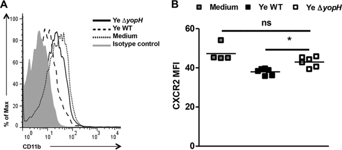
Expression of CXCR2 after in vitro infection. Blood neutrophils were in vitro infected with Y. enterocolitica WT or Y. enterocolitica ΔyopH at a multiplicity of infection (MOI) of 50:1 for 30 min. The cells were washed, and the CXCR2 expression in Ly6G+ CD11b+ cells was analyzed by flow cytometry. (A) Representative overlaid flow cytometry histogram analysis showing CXCR2 expression on neutrophil (Ly6G+ CD11b+) gate compared with uninfected cells (medium). Isotype control (gray peak). (B) The average CXCR2 mean fluorescence intensity (MFI) levels are indicated. The data are the summary results of 2 experiments. Each symbol represents cells from an individual mouse; horizontal lines indicate the means (*, P < 0.05; ns, not significant).