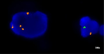
Fig. 1.

FISH on the urine cytology sample with probes for the X chromosome centromere (red) and the Y chromosome heterochromatic region (green) (Abbott Molecular, Downers Grove, IL); large malignant urothelial carcinoma cell with four X chromosome signals and a normal male cells with one X and one Y chromosome signal
