
Fig. 1.
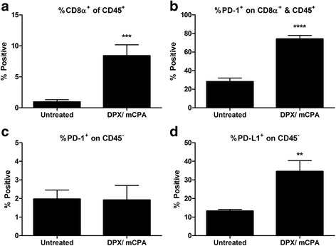
DPX vaccination and mCPA treatment increased infiltration of C3 tumors with PD-1+ CD8α+ T cells. Mice bearing C3 tumors were treated with mCPA and vaccinated with DPX-R9F. Eight days after vaccination, mice were terminated. Tumors were dissociated and analysed by flow cytometry for expression of CD45, CD8a, PD-1 and PD-L1. a Percent CD8α positive of CD45 positive cells; b Percent PD-1 positive of CD8α&CD45 double positive cells; c Percent PD-1 positive of CD45 negative cells; d Percent PD-L1 positive of CD45 negative cells Results pooled from two separate experiments, n = 6–8, average ± SEM, statistics by students t-test, ** p < 0.01, ***p < 0.0001