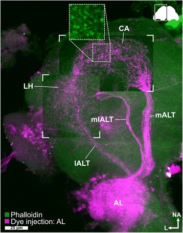
Fig. 5.

Antennal lobe tracts. Maximum intensity projection of a CLSM image stack after dye injection into the AL (magenta) revealed three antennal lobe tracts – the medial (mALT), mediolateral (mlALT), and the lateral antennal lobe tract (lALT) – as well as the calyx (CA) and the lateral horn (LH). In the CA, most fibers from the mALT form microglomeruli (inset obtained from another preparation). The staining in the optical lobe is an artifact caused by diffusion of the dye during application. Phalloidin counterstaining in green. AL antennal lobe, ALT antennal lobe tracts, CA calyx, CLSM confocal laser-scanning microscopy, lALT lateral antennal lobe tract, LH lateral horn, mALT mediolateral lobe tract, mlALT mediolateral lobe tract
