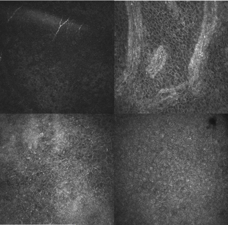
FIGURE 1.
Representative confocal images of a normal eye (top row) and total limbal stem cell deficiency eye (bottom row). The central cornea (top left) and limbus (top right) at the basal cell layer are shown in a normal eye. Distinct individual epithelial cells are observed in both the normal cornea and limbus. The central cornea (bottom left) and temporal limbus (bottom right) at the basal cell layer are shown in the total limbal stem cell deficiency eye. Few epithelial cells with borders and dark cytoplasm can be seen in the cornea. Only few visible cells with clear borders can be detected in the limbus.

