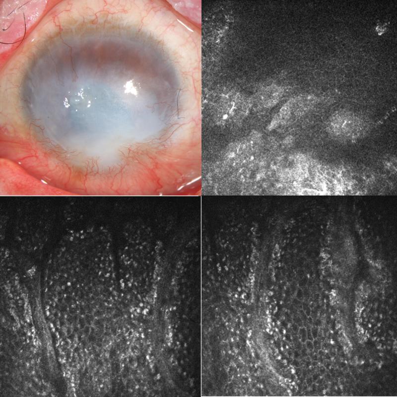
FIGURE 4.
Slit lamp photo of left eye of patient 3 (top left) shows 360 degree of superficial corneal neovascularization, inferior pannus and corneal scarring. Confocal images of the central cornea (top right), superior limbus (bottom left and bottom right) show the presence of normal basal limbal epithelial cells.

