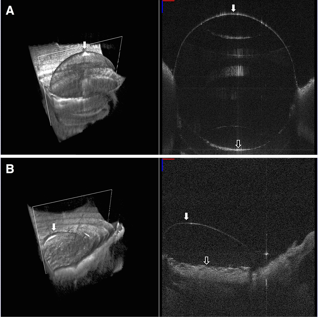
FIGURE 1. Endothelial side up swept-source microscope-integrated optical coherence tomography (SS-MIOCT) images after air injection to confirm big bubble (BB) formation result.
(Top) Volume (left) and B-scan (right) SS-MIOCT images from a full BB formation group sample showing a single, centrally located bubble >7 mm in diameter (white arrow). The separated corneal stroma (black arrow) could be easily visualized in the B-scan images. (Bottom) Volume (left) and B-scan (right) SS-MIOCT images from a partial BB formation group sample showing a single, peripherally located bubble <7 mm in diameter (white arrow).

