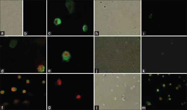
Figure 5.
Annexin-V/propidium iodide assay. In control cells propidium iodide cannot go into the cell resulting in negative staining for both dyes; (0 mg/ml after 4 and 8 h) (a, b, h and i, respectively), annexin-V stain externalized phosphatidylserine on the cell surface (green) next the beginning of apoptosis (1.25 mg/ml after 8 h) (j and k), through the latter stages of apoptosis phosphatidylserine is exposed (green) and propidium iodide can enter the cell (red) due to the loss of membrane integrity of late apoptotic cell (2.5 mg/ml (c) and 5 mg/ml (d-f) after 4 h, 2.5 mg/ml (l and m) after 8 and dead cell (5 mg/ml after 8 h) (g)

