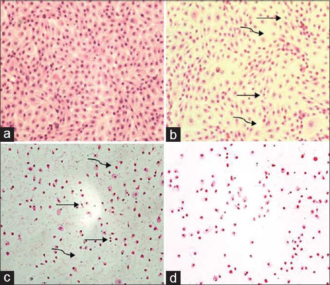
Figure 6.

H and E stain of Madin-Darby Bovine Kidney cells incubated for 8 h with khat extract; 0.0 mg/ml, Madin-Darby Bovine Kidney cells show complete confluent healthy monolayer cells (a), 1.5 mg/ml, very few cells show small, hyperchromic fragmented nuclei (curved arrow) and very few gaps (straight arrow) between cells (b), 2.5 mg/ml, many cells show small, hyperchromic, fragmented nuclei, many gaps between cells and cytoplasmic and nuclear dust were found (c) and in 5 mg all fields lack cells, with hyperchromic naked nuclei, cytoplasmic and nuclear stages of destruction, with detachment of most of the cells from adherent surface (d) (×100)
