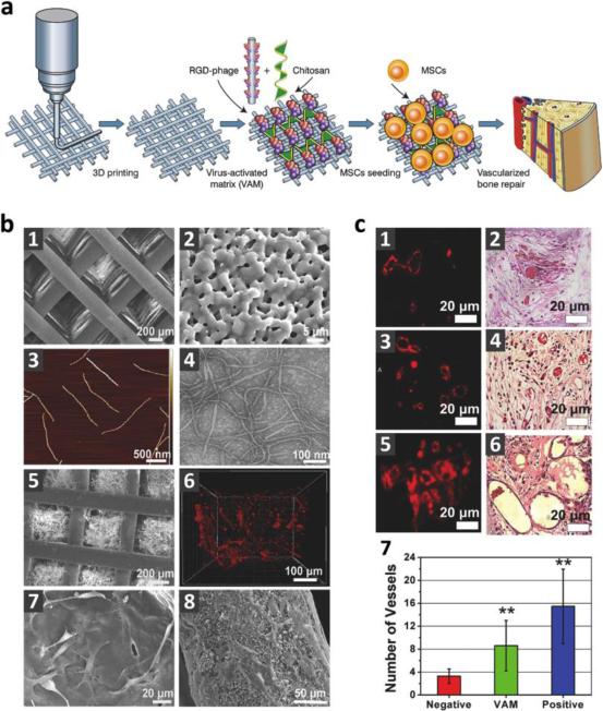
Figure 13. 3D printed virus-activated bone scaffold with angiogenesis.
a) Schematic of 3D printed bioceramic bone scaffold incorporating negatively charged RGD-labeled phage nanofibers using positively charged chitosan for new bone and blood formation when seeded with MSCs. b) Images of scaffold architecture. Scanning electron microscopy (SEM) of bone scaffold showed macro-scale (1) and micro-scale (2) pores, as well as pores filled with chitosan and phage matrix (5). Atomic force microscopy (AFM) (3) and transmission electron microscopy (TEM) (4) demonstrated morphology of phage nanofibers. 3D confocal fluorescence imaging showed presence of dye-labeled phage (red) within matrix-filled pores (6), and brightfield imaging revealed support of MSC adhesion for both the scaffold pores (7) and columns (8). c) Immunofluorescence staining for endothelial CD31 (1, 3, 5) and hematoxylin and eosin (H&E) staining (2, 4, 6) of implants of negative control (wild-type phage), virus-activated matrix (VAM), and positive control (RGD-phage with VEGF) scaffolds, respectively, as well as quantitative analysis (7) showed VAM promotes angiogenesis at an intermediate level. (**p<0.01). Reproduced with permission from ref. 334. Copyright 2014 John Wiley & Sons.

