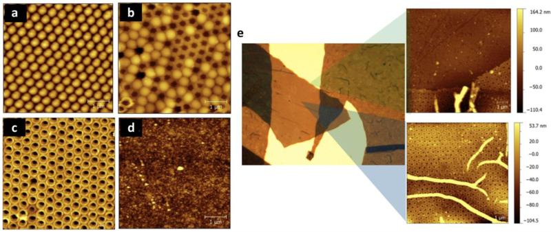
Figure 23. Creation of free-standing Janus mesoporous virus film.
Topographical tapping mode AFM images illustrate the initial formation of close-packed PS microspheres (a), partial removal of PS spheres (b) after patterned poly(pyrrole-co-pyrrole-3-carboxylic acid) electropolymerization (c), and a non-patterned film (d). e) Optical microscope image (480 μm by 360 μm) after overlaying CPMV on the patterned polymer through electrostatics and delaminating the film to create a free-standing film. Insets show topographical AFM images at indicated points in the film. Reproduced with permission from ref. 538.

