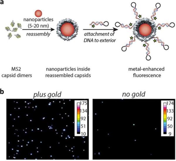
Figure 26. Controlled display of gold nanoparticles and fluorophores for enhanced fluorescence.
a) Schematic for the assembly of MS2 around gold nanoparticles followed by attachment of DNA hairpins to place fluorophores a fixed distance away from the capsid. b) Images from total internal reflection fluorescence microscopy of MS2 labeled with fluorophores set 3 bp away from the capsid, with (left) and without (right) gold encapsulated, demonstrating metal-enhanced fluorescence. Reproduced with permission from ref. 174. Copyright 2013 American Chemical Society.

