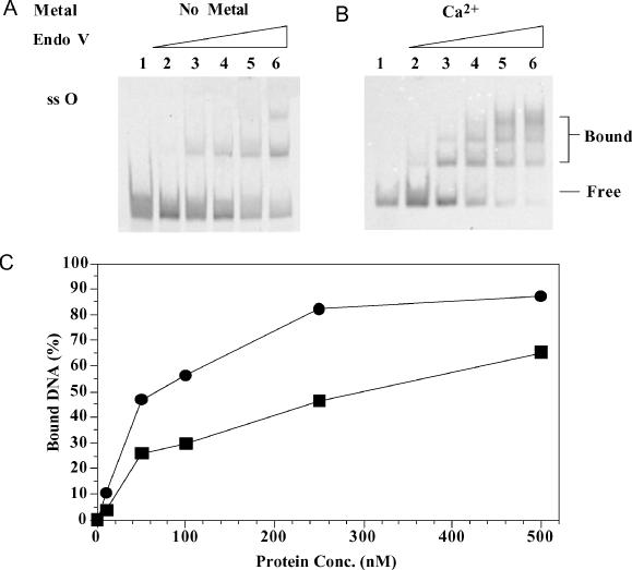
Figure 7.

Gel mobility shift analysis of Salmonella endo V with single-stranded O-containing substrate. Lanes: 1, 0 nM endo V; 2, 10 nM endo V; 3, 50 nM endo V; 4, 100 nM endo V; 5, 250 nM endo V; and 6, 500 nM endo V. (A) Binding of single-stranded O-containing substrate without metal. (B) Binding of single-stranded O-containing substrate with Ca2+. (C) Quantitative analysis of endo V binding to single-stranded O-containing substrate without metal (squares) or with Ca2+ (circles).
