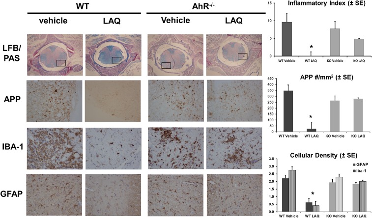
Fig. 5.
Prophylactic treatment with laquinimod has no effect on inflammatory demyelination, acute axonal damage, or microglial and astroglial activation in AhR−/− mice. All stains were performed using n = 15 per treatment group. The inflammatory index was quantified using LFB/PAS and H&E staining of spinal cords taken from vehicle- or laquinimod-treated WT or AhR−/− mice. (Upper panels) A cross-section of the whole spinal cord using 4× objective (Nikon Eclipse E200). (Lower panels) A higher magnification (40×) taken from the area marked by a rectangle. Quantification of acute axonal damage using APP staining showed a significant reduction (*P < 0.0001) in the number of APP+ spheroids in laquinimod-treated WT mice, but not in AhR−/− mice. Quantification of microglial and astrocyte activation using Iba-1 or GFAP staining showed a significant reduction (*P < 0.0001) in the number of Iba-1 (black bars) and GFAP (striped bars) cells in laquinimod-treated WT mice, but not in AhR−/− mice.

