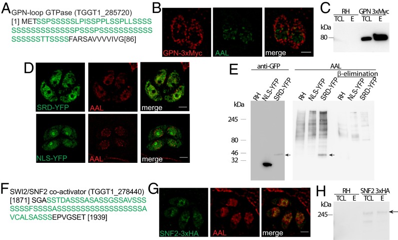
Fig. 4.
O-fucosylation directs proteins to the nuclear periphery. (A) The N-terminal SRD of GPN. (B) An ectopic copy of the protein localizes to the cytoplasm and to the nuclear periphery with AAL by ELYRA SIM. (C) AAL pull-down followed by anti–c-MYC Western blotting shows GPN enrichment. (D) SIM shows that the GPN SRD fused to YFP (SRD-YFP) partially colocalizes with AAL, but NLS-YFP does not. (E) AAL recognizes an additional band only in the SRD-YFP cell lysate, consistent with the molecular weight of SRD-YFP as defined by anti-GFP blot (black arrows). (F) The SRD of the SNF2 transcriptional coactivator. (G) SNF2 colocalizes with AAL at the nuclear periphery. (H) AAL pull-down followed by anti-HA blotting shows that SNF2 is present in the AAL-bound fraction (black arrow). E, elution; RH, wild-type cell lysate; TCL, total cell lysate. (Scale bars: 2 μm.)

