Fig. 1.
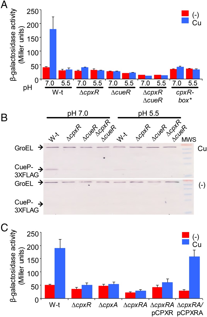
Copper-induced expression of cueP depends on CpxR. (A) β-gal activity from a cueP::lacZ transcriptional fusion expressed on wild-type (W-t), ΔcpxR, ΔcueR, ΔcpxR ΔcueR, and cpxR-box* cells grown overnight in LB broth at pH 7.0 or pH 5.5 and without (−) or with the addition of 1 mM CuSO4 (Cu). The data correspond to mean values of four independent experiments performed in duplicate. Error bars represent SD. (B) Analysis of the expression of CueP-FLAG, using anti-FLAG antibodies. Here 20 μg of total crude extract protein of wild-type, ΔcpxR, ΔcueR, and ΔcpxR ΔcueR cells grown overnight in LB broth at pH 7.0 or pH 5.5 and without (−) or with (Cu) the addition of 1 mM CuSO4 was analyzed by SDS/PAGE, followed by transfer to nitrocellulose and development using monoclonal anti-FLAG antibodies. The PageRuler prestained protein ladder provided molecular weight standards. From top to bottom, bands of 70, 55, 40, 35, 25, and 15 kDa are shown. (C) β-Gal activity from the cueP::lacZ transcriptional fusion expressed on wild-type, ΔcpxR, ΔcpxA, ΔcpxRA, and cpxRA strains complemented with pCPXR (ΔcpxR/pCPXR) or with pCPXRA (ΔcpxR/pCPXRA) without (−) or with the addition of 1 mM CuSO4 (Cu). The data correspond to mean values of four independent experiments performed in duplicate. Error bars represent SD.

