Fig. 4.
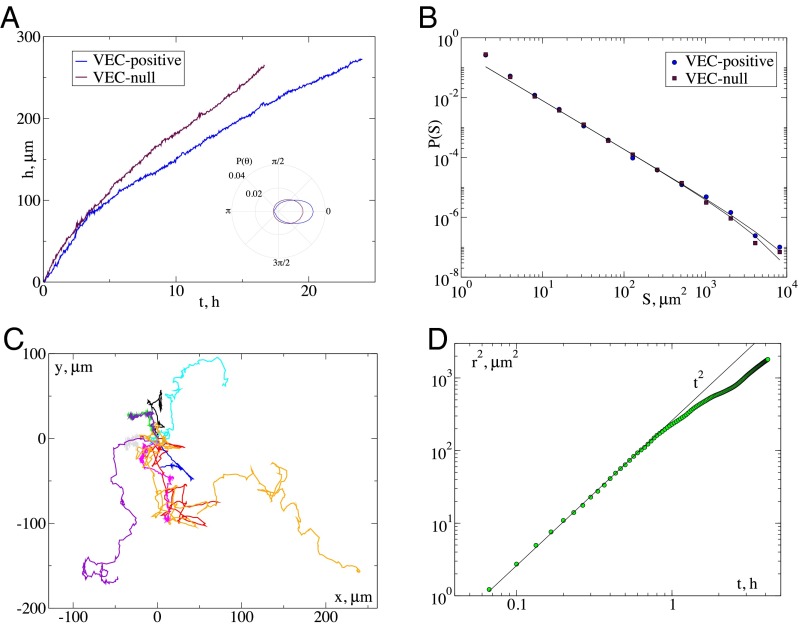
Knockdown of VE-cadherin leads to faster fronts and individual cell motion. (A) The time evolution of the front position for mouse endothelial cell expressing (VEC-positive) or not (VEC-null) VE-cadherin. VEC-null cells move faster. (B) The corresponding cluster size distribution displays the same exponent and small changes in the cutoff. (C) In the VEC-null cases cells detach from the front and invade the space individually following trajectories as the ones illustrated. (D) The average mean-square displacement of the trajectories indicates an initial ballistic regime followed by a slowing down.

