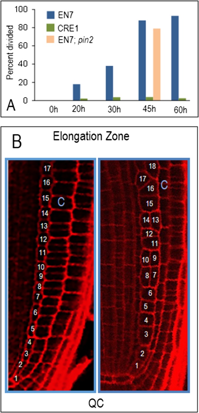
Fig. S1.
Loss of PD signaling to and from the endodermis but not the stele affects the orientation of cell divisions in the endodermis. (A) Quantification of cells within an endodermal file that had divided periclinally. One hundred percent would mean that all cells in the endodermal file had undergone at least one periclinal cell division. EN7 and CRE1 represent EN7:icals3m and CRE1:icals3m, respectively; EN7;pin2 represents EN7:icals3m in the pin2 background. (B) Diagram showing how the number of cells in the endodermal lineage was counted. All cells in the endodermal cell file from the QC to the elongation zone were counted.

