Fig. 8.
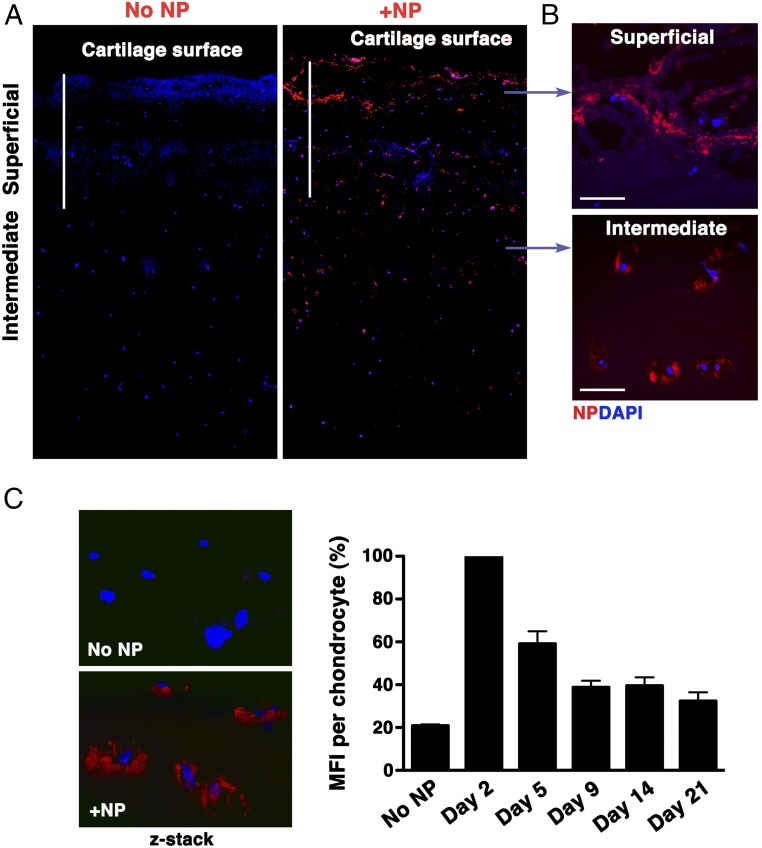
NP penetration in human OA cartilage explants. p5RHH-Cy3–labeled siRNA NPs (red) were incubated with 5-mm2 cartilage explants from human OA knee. (A) After 48 h, cartilage explants were washed extensively and processed for histology. Sagittal sections were examined for depth of NP penetration by confocal microscopy. DAPI (blue) stains nuclei. (B) Large aggregates of NPs could be seen in the matrix of the superficial zone and accumulated in chondrocytes in the intermediate zone. (C) Human OA cartilage explants were incubated with fluorescent NPs for 48 h; the excess NP was washed off, and explants were kept in complete culture medium for up to 21 d. Fluorescent signal inside chondrocytes was examined by confocal microscopy at different time points. MFI per chondrocyte was obtained from z-stack confocal images (at least 20 chondrocytes per time point were analyzed); data were derived from two different OA samples. The rate of fluorescence decay was calculated relative to the signal observed on day 2, which was set at 100%. [Scale bars, 500 μm (A) and 50 μm (B).]

