Fig. S2.
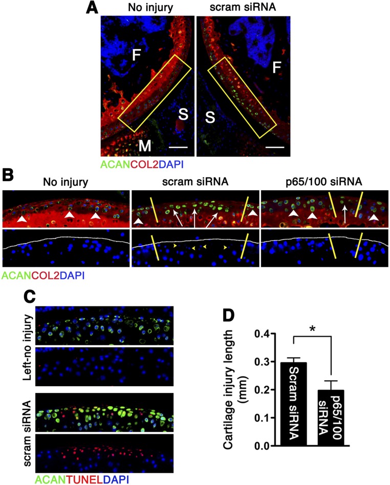
Aggrecan redistribution following impact injury and p5RHH-p65/p100 siRNA NP administration. Mouse knees were subjected to compression injury at 6 N and injected i.a. immediately and at 48 h with 0.1 μg of p5RHH-p65/p100 siRNA combined or scrambled (scram) siRNA NP. On day 5, knees were processed and examined for aggrecan (ACAN) distribution. (A) Mechanical loading changed the distribution pattern of ACAN in the impact area. (Scale bars, 100 μm.) (B) Higher magnification of the impact area (rectangles in A) revealed redistribution of ACAN from pericellular (white arrowheads) to intracellular (white arrows) in the injured area (demarcated by yellow lines). Cells with ACAN redistribution had pyknotic nuclei (yellow arrowheads) that stained poorly with DAPI. (C) Cells with ACAN redistribution also correlated with TUNEL+ cells. (D) Length of cartilage lesion (based on ACAN redistribution) in p5RHH-scram siRNA vs. p5RHH-p65/p100 siRNA NP treatment. Values represent mean ± SEM. n = 4 mice per treatment group. COL2 (red), type II collagen; F, femur; M, meniscus; S, synovium. DAPI (blue) stains nuclei. *P < 0.05.

