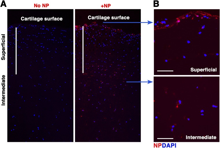
Fig. S7.
p5RHH-siRNA NP penetration of normal cartilage. Normal human cartilage discs were incubated with Cy3-labeled NP for 48 h after which the excess NP was washed off. Presence of fluorescent signal in cartilage was examined by confocal microscopy. DAPI (blue) stains nuclei. (A) NP can be seen diffusing freely into the intermediate zone of cartilage. (B) NP accumulates at the cartilage surface and inside chondrocytes located in the deeper zone. [Scale bars, 500 μm (A) and 50 μm (B).]

