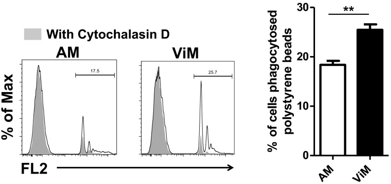
Fig. S9.
Phagocytosis of latex beads by the macrophages. Lung macrophages isolated from immunized plus CY-treated mice were incubated in vitro with fluorescent latex beads for 60 min. Representative flow data (Left) and the fluorescence-positive fractions in macrophage gates (Right) are shown. Bars represent mean of three to four replicates from an experiment using macrophages pooled from 15 mice. Error bars represent SEM. **P < 0.01 by unpaired t test. FL2, fluorescence channel 2.

