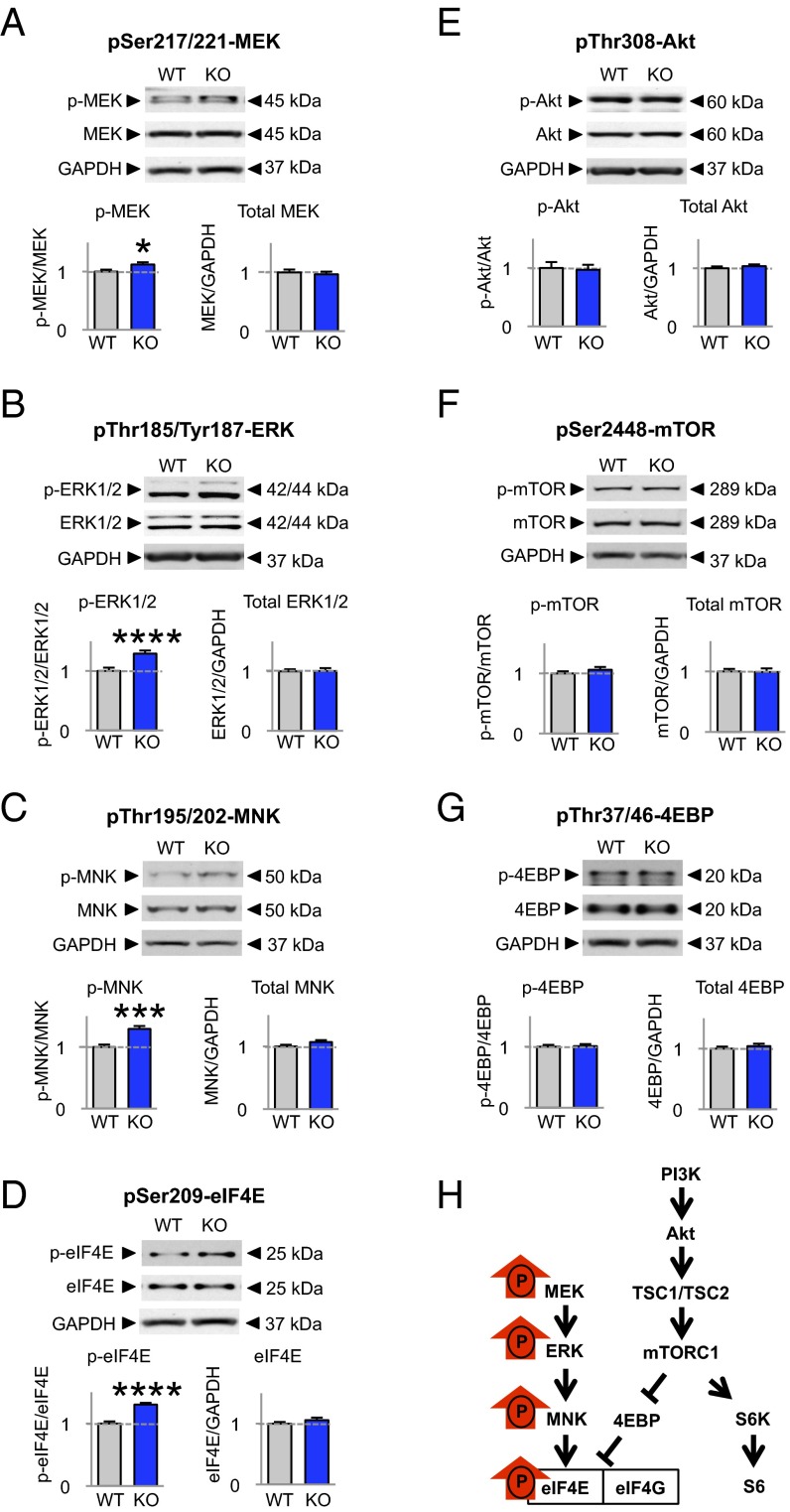
Fig. 1.
MEK/ERK signaling, but not PI3K/mTOR signaling, is dysregulated in the neocortex of Fmr1 KO mice. (A–G) Representative Western blots of cortical lysates from 4-wk-old WT and Fmr1 KO mice and summary data showing relative abundance of phosphorylated and total protein. Western blots were probed for proteins in the ERK and mTOR pathways. Phosphorylation (but not total abundance) was elevated for proteins in the MEK/ERK pathway: p-MEK (WT, n = 7; KO, n = 9) (A), p-ERK (WT, n = 22; KO, n = 24) (B), p-MNK (WT, n = 19; KO, n = 16) (C), and p-eIF4E (WT, n = 18; KO, n = 16) (D). In contrast, there was no detectable difference in the phosphorylation status of proteins in the mTOR pathway: p-Akt (WT, n = 5; KO, n = 6) (E), p-mTOR (WT, n = 7; KO, n = 9) (F), and p-4EBP (WT, n = 17; KO, n = 16) (G). Data represent mean ± SEM; *P < 0.05; ***P < 0.001; ****P < 0.0001. (H) Schematic of ERK and mTOR signaling in the neocortex of Fmr1 KO mice depicting proteins with elevated basal phosphorylation (red arrows).

