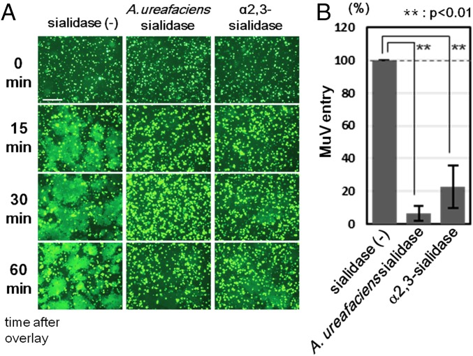
Fig. 4.
Effect of cleavage of sialic acid on MuV-induced cell–cell fusion and MuV entry. (A) HEK293 cells expressing EGFP were treated with control medium, α2,3-sialidase, or A. ureafaciens sialidase. They were detached from the plates and then overlaid onto HEK293 cells expressing the HN and F proteins of MuV. The cells were observed under fluorescence microscopy at 0, 15, 30, and 60 min after overlay. (Scale bar: 200 μm.) (B) HEK293 cells pretreated with control medium, α2,3-sialidase, or A. ureafaciens sialidase were infected with the EGFP-expressing recombinant MuV. At 24 h postinfection, EGFP-positive cells were counted to evaluate the efficiency of virus entry. The control was set to 100, and data indicate the mean ± SD of triplicate experiments. The data are representative of three independently performed experiments. **P < 0.01, two-tailed Student’s t test.

