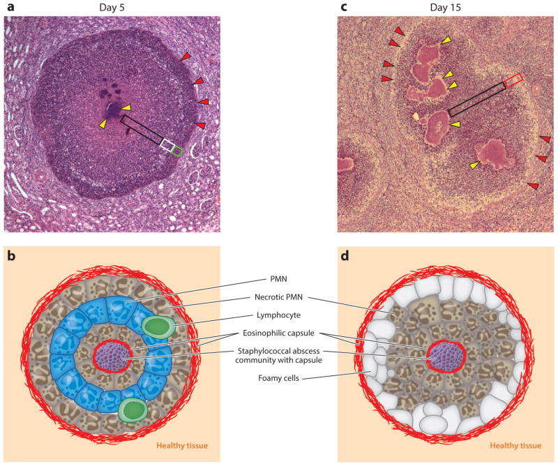
Figure 3.
Staphylococcus aureus abscess lesions in murine renal tissue. (a) Microscopy image of hematoxylin-eosin-stained, thin-sectioned renal tissue 5 days following intravenous inoculation with S. aureus. Bacteria are organized into staphylococcal abscess communities (SACs) ( yellow arrowheads) at the center of the lesion. Depending on sectioning, SACs may appear fragmented; however, bacterial communities remain connected and are enclosed by eosinophilic deposits of fibrin and surrounded by zones of necrotic neutrophils (black box), healthy-appearing neutrophils (white box), and necrotic immune cells ( green box). Abscess lesions are demarcated from renal tissues by another layer of eosinophilic deposits (red arrowheads), through which immune cells enter the lesion. (b) Schematic illustrating the histopathology features of a 5-day-old S. aureus abscess lesion. (c) Microscopy image of hematoxylin-eosin-stained, thin-sectioned renal tissue 15 days following intravenous inoculation with S. aureus. The 15-day-old lesion differs from the 5-day-old lesion in that SACs have expanded and the layering of healthy and necrotic immune cells has been supplanted by diffuse infiltrates of immune cells, cellular detritus, liquefaction (black box), and foam cells positioned at the periphery of the lesion (red box). SACs remain enclosed by fibrin deposits ( yellow arrowheads). An outer eosinophilic layer (red arrowheads) demarcates the lesion from large numbers of immune cells and from the cellular detritus that has replaced renal tissue architecture in the vicinity of the abscess lesion. (d ) Schematic illustrating the histopathology features of a 15-day-old S. aureus abscess lesion. Abbreviation: PMN, polymorphonuclear leukocyte.

