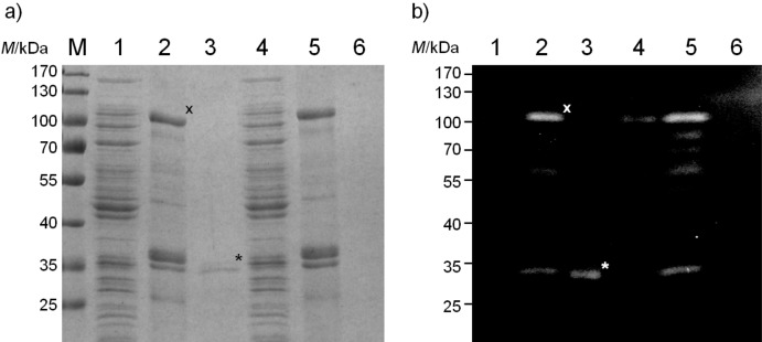
Fig. 10.
Differential cell fractionation of E. coli cells expressing MATE-mCherry from either an OmpT-positive strain UT2300 (lanes 1–3) or an OmpT-negative strain UT5600 (lanes 4–6). a) SDS-PAGE stained with Coomassie Brilliant Blue; b) Western blot with antibody against 6xHis epitope. M=PageRuler prestained protein marker. Fractions of E. coli soluble proteins (lanes 1 and 4), outer membrane proteins (lanes 2 and 5) and secreted proteins purified from the growth medium (lanes 3 and 6) are shown. Sample loading was normalized so that the proteins from an equivalent number of cells is given in each lane. The MATE-mCherry fusion protein is marked with an x, the secreted mCherry is marked with an asterisk (*)

