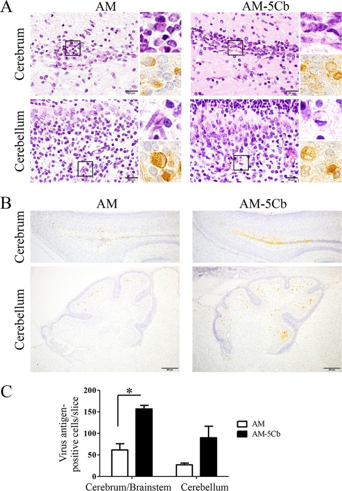
FIG 6.

Histopathological results and viral antigens in the brains of neonatal mice after intracerebral inoculation with mouse-passaged SAFV-3. Neonatal ddY mice were intracerebrally inoculated with the AM or AM-5Cb strain at 104 CCID50s/10 μl. (A) The areas of the cerebral ventricles and cerebellum of a neonatal mouse after infection. Top left and right, H&E staining; bottom right, immunohistochemical staining with an anti-SAFV antibody. Degenerated neural cells are present in the cerebral ventricles and cerebella of AM- and AM-5Cb-inoculated mice. The degenerated neural cells with eccentric nuclei are positive for viral antigens (insets). (B) Viral antigen-positive cells are located mainly in the areas of the cerebral ventricles and cerebella of AM- and AM-5Cb-inoculated mouse strains. Original magnifications, ×1,000 (panel A and all insets) and ×100 (B). Bar, 20 μm (left side of panel A) and 200 μm (B). (C) Viral antigen-positive cells were counted in two slices per mouse (n = 3 each group). Numbers of viral antigen-positive cells in the cerebrum/brainstem and cerebellum were compared. *, P < 0.05; unpaired t tests.
