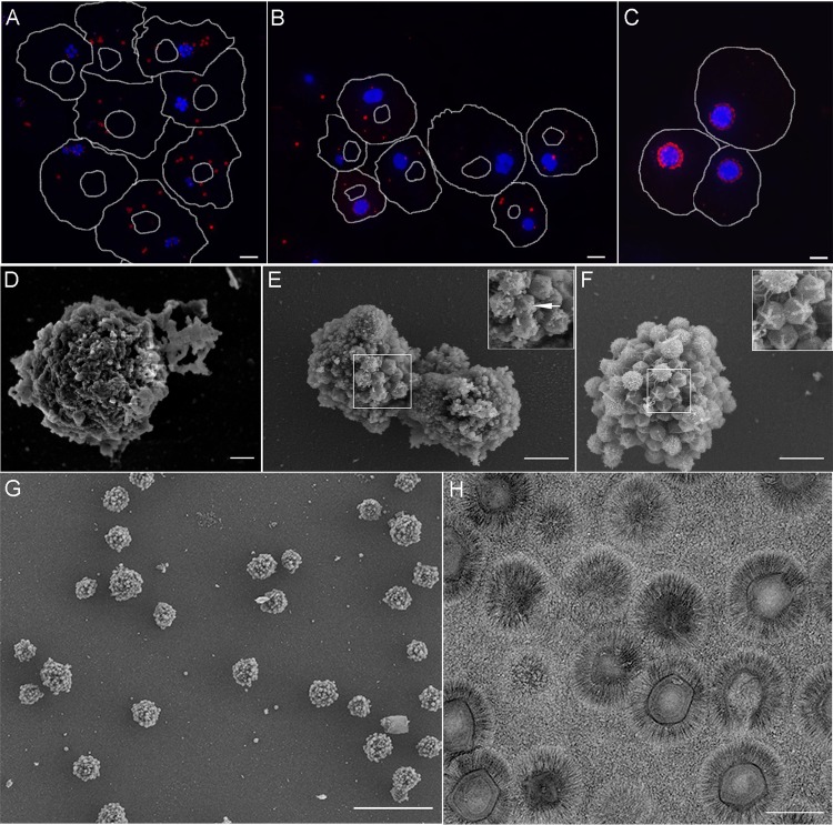
FIG 1.
Mimivirus factories within host cells and following isolation. (A to C) Amoeba cells infected with mimivirus at 3 successive postinfection (p.i.) time points (4 h [A], 5.5 h [B], and 7 h [C]) were stained with antibodies against mature virions (red) and counterstained with DAPI (blue). Cell and nucleus contours were derived from differential interference contrast (DIC) micrographs. (D to F) SEM images of isolated viral factories at 4 h (D), 5.5 h (E), and 7 h (F) p.i. Both SEM and fluorescence studies revealed the coalescence of initial viral replication centers into a single viral factory. Insets in panels E and F show stargate structures. (G) Low-magnification SEM micrograph depicting a large field of isolated factories at 7 h p.i. The micrograph reveals that the factories are essentially free from large host components. (H) Purified virions appear pure when surveyed by low-magnification TEM. Scale bars: panels A to C, 5 μm; panel D, 200 nm; panels E to F, 1 μm; panel G, 10 μm; panel H, 500 nm.

