
FIG 3.
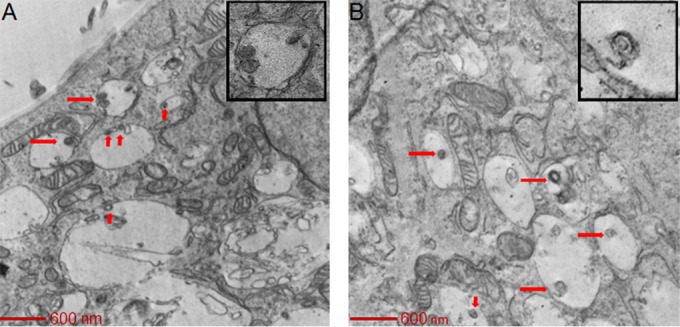
HCMV virions enter SNB-19 cells via macropinocytosis. HCMV was added to SNB-19 cells on ice for 60 min and then internalized for 90 min and observed under a transmission electron microscope as described in the legend to Fig. 2. In 20 electron microscopy fields with virus-infected SNB-19 cells, 87% (33/38) of HCMV particles were inside macropinosomes, while 13% (5/38) of HCMV particles were free in the cytoplasm.