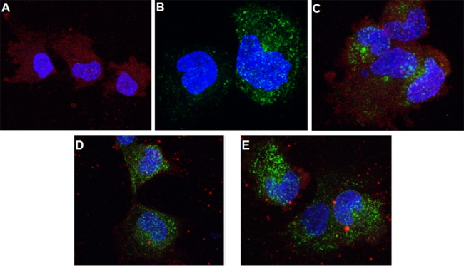
FIG 4.
HCMV internalization does not involve clathrin-coated endosomes. The results of a confocal microscopy study using a Leica SP5 microscope are shown. (A) HS-578T cells were exposed to HCMV on ice, followed by internalization at 37°C for 45 min, and virus internalization was terminated using a low-pH citrate buffer wash to inactivate noninternalized virus and remove the soluble proteins. The cells were then fixed and stained with monoclonal anti-pp65 followed by anti-mouse–Alexa 594 (red) secondary antibody. Nuclei were stained with DAPI (4′,6′-diamidino-2-phenylindole). (B) HS-578T cells were incubated with transferrin-conjugated Alexa 488 (green) on ice and then at 37°C for 45 min. After a low-pH buffer wash as described above, the cells were fixed and observed under the microscope. (C) HS-578T cells were incubated with both transferrin and HCMV as described for panels A and B. After the acid buffer wash, the cells were fixed and stained in the same manner as cells shown in panel A. (D and E) HS-578T cells were incubated with transferrin alone (D) or with both transferrin and HCMV (E) as described for panel C and then fixed and stained with a goat anti-THY-1 antibody followed by anti-goat–Alexa 549 (red) secondary antibody.

