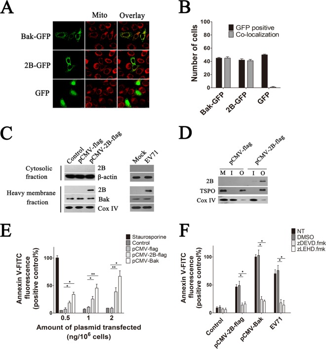
FIG 1.
(A) Confocal microscopy analysis of the colocalization of 2B-GFP and Bak-GFP within the mitochondria in HeLa cells. HeLa cells were transfected with pGFP, pGFP-2B, or pGFP-Bak. At 24 h posttransfection, they were stained with MitoTracker Red and subjected to confocal microscopy analysis. An overlay of the MitoTracker Red (red) and GFP florescence (green) is also shown. Mito, mitochondria. (B) Colocalization index estimation for cells transfected with pGFP, pGFP-2B, or pGFP-Bak and labeled for MitoTracker Red as for panel A. To determine the percentage of colocalization, green and merged images were loaded into the Leica TCS SP8 platform, and the ratio of GFP-positive cells to counted cells (in a total of 100 cells) was determined. Cells with GFP and mitochondrion colocalization (Manders overlap coefficient of >0.8) were counted. (C) Immunoblotting analysis of 2B-Flag in the cytosol and mitochondrial fractions of HeLa cells infected with EV71 or transfected with pCMV-2B-Flag. Cytosolic and heavy membrane fractions were separated as described in Materials and Methods. Mitochondrial fractions were processed for immunoblotting with antibodies specific to EV71 2B, Bak, and Cox IV. Cytosolic fractions were immunoblotted with antibodies to EV71 2B and β-actin (internal control). Control, cells transfected with Tris-acetate-EDTA (TAE) buffer. Mock, cells with no EV71 infection. (D) Western blot analysis of the sublocation of 2B. Cells were transfected with pCMV-2B-Flag or pCMV-Flag. The mitochondrial fractions were isolated at 48 h posttransfection. The inner and outer mitochondrial membranes were isolated by sucrose gradient centrifugation and immunoblotted with an antibody to the Flag tag, Cox IV, and TSPO (internal control of outer mitochondrial membrane fraction). M, mitochondria of cells transfected with pCMV-Flag. I, inner mitochondrial membrane fraction. O, outer mitochondrial membrane fraction. (E) Analysis of cell apoptosis induced by 2B. Cells were treated with 30 nM staurosporine (positive control) or transfected with increasing amounts of pCMV-2B-Flag, pCMV-Flag, or pCMV-Bak. At 24 h posttransfection, cells were harvested and stained with annexin V-FITC. The percentages of apoptotic cells corresponding to the respective fluorescence intensities were calculated. (F) Analysis of the antiapoptosis effects of caspase inhibitors on 2B-induced apoptosis. Cells either infected with EV71 or transfected with pCMV-2B-Flag or pCMV-Bak-Flag (positive control) were treated with dimethyl sulfoxide (DMSO), zDEVD.fmk, or zLEHD.fmk for 24 h and then stained with annexin V-FITC and subjected to flow cytometry. NT, cells with no treatment. Control, cells transfected with pCMV-Flag.

