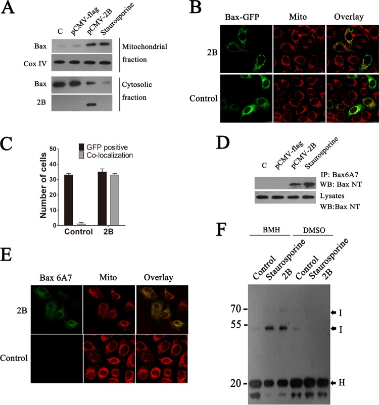
FIG 4.
Effects of EV71 2B on the translocation and activation of Bax. (A) Western blot analysis of Bax and activated Bax in the cytosol and mitochondrial fractions of cells transfected with pCMV-Flag or pCMV-2B-Flag. Cytosolic and mitochondrial fractions were separated, and equal amounts of proteins from each fraction were immunoblotted with anti-BaxNT, anti-Flag, or anti-Cox IV (internal control of mitochondrial fraction). Control, cells transfected with TAE buffer. Staurosporine, cells treated with staurosporine (30 nM). (B) An experiment indicating that 2B induces the translocation of Bax to the mitochondria. Cells were cotransfected with pGFP-Bax (green) and pCMV-Flag (control) or pGFP-Bax and pCMV-2B-Flag. At 12 h posttransfection, the cells were fixed and analyzed by confocal fluorescence microscopy. Red, MitoTracker Red. An overlay of the MitoTracker Red and GFP florescence is also shown (merged). (C) Colocalization indexes. Cells were cotransfected with pGFP-Bax and pCMV-Flag or pCMV-2B-Flag. At 24 h posttransfection, cells were labeled with MitoTracker Red and examined under a confocal laser scanning microscope. Cells with GFP and mitochondrial colocalization (Manders overlap coefficient of >0.8) were counted as described for Fig. 1. (D) Western blot analysis of the Bax activation in cells transfected with the same panel of pCMV-Flag or pCMV-2B-Flag plasmids. Cells were lysed and immunoprecipitated with anti-Bax6A7 antibody. Equal amounts of the precipitated protein and cell lysates were immunoblotted with antibody to Bax. (E) Immunofluorescence analysis of the Bax activation in cells transfected with pCMV-2B-Flag (2B). The cells were incubated with MitoTracker Red (red) for 10 min, fixed, and stained with anti-Bax6A7 (green). (F) An experiment indicating that 2B induces homo-oligomerization of Bax. Mitochondria from cells transfected with pCMV-Flag or pCMV-2B-Flag were cross-linked with BMH or DMSO as described in Materials and Methods. Equal amounts of proteins from each sample were immunoblotted with anti-Bax antibody. H, Bax homo-oligomers; I, monomeric intramolecularly cross-linked Bax species.

