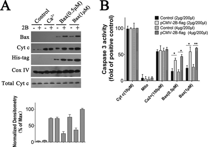
FIG 6.

Experiments showing that Bax induces the release of Cyt c from isolated mitochondria in the presence of 2B. (A) Western blot analysis of the impact of recombinant Bax on Cyt c release in isolated mitochondria. The mitochondria were incubated for 1.5 h with either 1 μM recombinant Bax, 0.5 μM recombinant Bax, 150 μM Ca2+, or none of these reagents (control) at RT. They were then pelleted by centrifugation, and the resulting supernatants were immunoblotted with antibody to Cyt c. The pellet fractions were lysed and immunoblotted with antibody to His tag or Bax. Inputs of equivalent amounts of mitochondria and total Cyt c from the mitochondria were verified by immunoblot analysis of the mitochondrial fractions using an antibody to Cox IV or Cyt c, respectively. Densitometry of the Cyt c band normalized to Cox IV is presented as fold change compared with Cyt c release in isolated mitochondria incubated with 1 μM recombinant Bax, defined as 100. (B) Experiments showing that Bax induces the release of mitochondrial factors which trigger the processing and activation of cytosolic caspase-3 in mitochondria expressing 2B. Cells were transfected with 2 μg or 4 μg of pCMV-Flag or pCMV-2B-Flag. At 24 h posttransfection, mitochondrial fractions were separated as described in Materials and Methods. Mitochondria (Mito) or supernatants (400 μl) from mitochondria that had been incubated with either Bax or Ca2+ (150 μM) were incubated with purified cytosol (100 μl) at 25°C for 1.5 h. Aliquots were then analyzed for caspase-3 activity as described above. Positive control, purified cytosol treated with Cyt c (10 μM).
