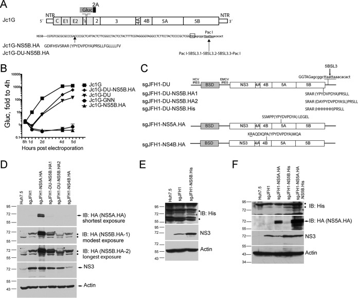
FIG 1.
Tagging of HCV nonstructural proteins in the context of viral replication. (A) Schematic of HCV Jc1G constructs. Duplicated 5BSL3 is inserted into the artificially introduced PacI site in Jc1G-DU. The stop codon of NS5B (boxed) and the artificially introduced PacI site (underlined) are shown. The HA peptide sequence (bracketed) is inserted into the NS5B protein sequence. Gluc, Gaussia luciferase; NTR, UTR; 2A (black bar), the foot-and-mouth disease virus 2A autoproteolytic peptide. (B) Huh7.5 cells were transfected with in vitro-transcribed RNAs. Luciferase activity in supernatants was measured at various times and normalized to the value obtained at 4 h. Mean values ± standard deviations are shown (n = 3). Gluc, relative light units. Similar results were obtained in multiple independent experiments. (C) Schematic of HCV sgJFH1 constructs. Peptide sequences inserted into viral nonstructural proteins are shown in parentheses. (D) Western blotting (immunoblotting [IB])of pooled replicon cells with the antibodies indicated. Arrows indicate the specific protein bands. Dots indicate nonspecific bands. The asterisks indicate specific NS5B-HA bands. (E) Western blotting of sgJFH1-NS5B.His replicon cells. Dots indicate nonspecific bands. The asterisk indicates a specific NS5B-His band. (F) Western blotting of sgJFH1-NS5A.HA.NS5B.His replicon cells. The arrow indicates specific NS5A-HA bands. Dots indicate nonspecific bands. The asterisk indicates a specific NS5B-His band. The migration of size standards (in kilodaltons) in SDS-PAGE is indicated to the left of the gels in panels D to F.

