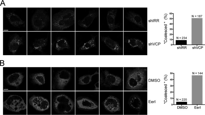
FIG 7.
Abolishing VCP function resulted in aberrant HCV NS5A distribution. (A) Huh7 cells were transduced with shIRR and shVCP lentiviruses. Two day later, the cells were cotransfected with phCMV-3-5B.ypet and VCP-es-DN at a ratio of 1:1 (microgram per microgram). At 1 day posttransfection, the cells were fixed and observed by confocal microscopy. The cells with a “coalesced” phenotype were enumerated, and the numbers were plotted (N is the total number of cells enumerated). Representative images are shown. (B) Huh7 cells were transfected with phCMV-3-5B.ypet. At 1 day posttransfection, the cells were treated with 2 μM EerI. One day later, the cells were fixed and observed by confocal microscopy. The cells with a “coalesced” phenotype were enumerated, and the numbers were plotted (N is the total number of cells enumerated). Representative images are shown. Scale bars, 10 μm. DMSO, dimethyl sulfoxide.

