FIG 2.
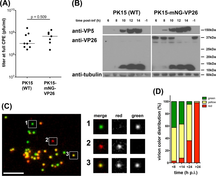
PK15-mNG-VP26 cells produce dual-color PRV180G. (A) Titer of PRV stocks grown on wild-type (WT) PK15 (n = 9) and PK15-mNG-VP26 (n = 7) cells until full CPE indicates no difference (P = 0.509) in maximal titers of either cell line. (B) Protein levels in lysates of PK15 (wild-type) and PK15-mNG-VP26 cells infected with PRV Becker (untagged VP26) for the indicated times were evaluated by immunoblotting using anti-VP26 (small capsid protein), anti-VP5 (large capsid protein), and anti-tubulin antibodies. Synthesis of untagged VP26 was detectable at 8 to 10 h p.i., together with VP5 expression. At 10 to 12 h p.i., mNG-VP26 expression declined rapidly. (C) Micrograph of purified PRV180G capsids harvested at 8 h p.i., spotted onto a coverslip, and imaged at a ×100 magnification in red and green channels. Three individual capsids were chosen to illustrate green, red, and yellow color profiles. Bar, 5 μm. (D) Bar graph indicating the color profile of PRV180G depending on the harvest time. PK15-mNG-VP26 cells were infected with PRV180 at an MOI of 1 before supernatants were collected at the indicated times p.i., and the color of >750 particles per stock was analyzed.

