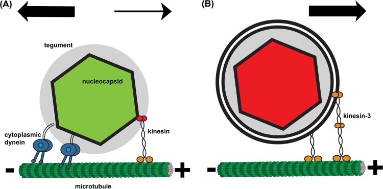
FIG 7.
Cartoon illustration of axonal PRV capsid transport. (A) During entry, green PRV180G capsids are transported by cytoplasmic dynein, which binds to the tegument layer (gray), and kinesin. The overall net transport is toward the microtubule minus end. (B) During egress, red PRV180G capsids are contained in a vesicle, which is transported only by kinesin motors (kinesin-3 and potentially others) toward the microtubule plus end.

