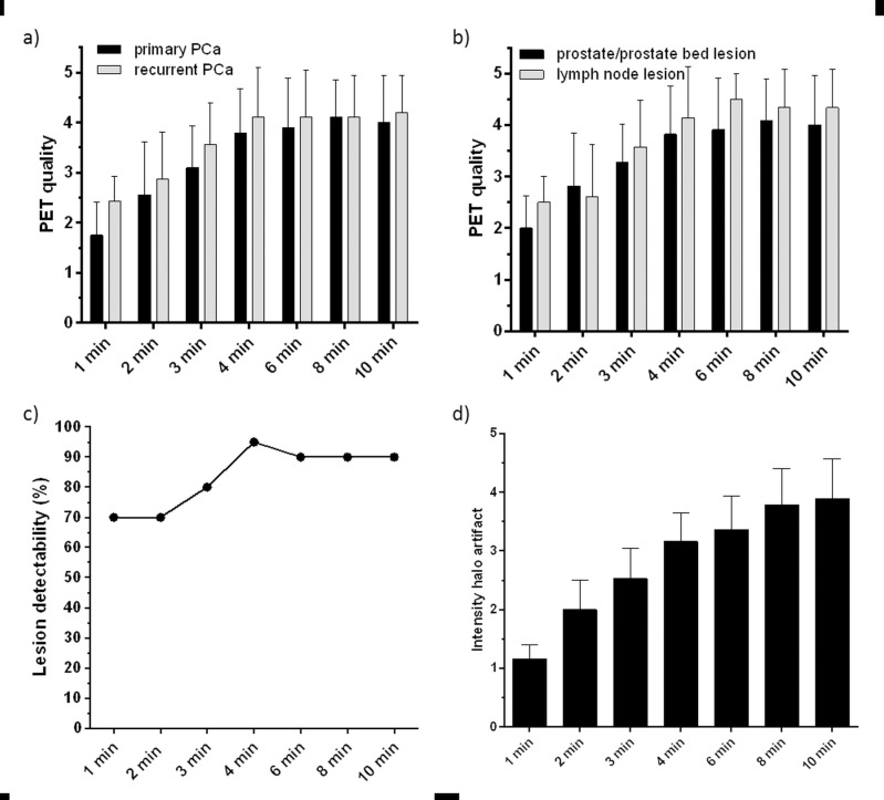
Fig 1.
Quality of PET images obtained with 68Ga-HBED-CC-PSMA PET/MRI at different acquisition times for primary (n = 10) and recurrent (n = 10) PCa lesions (a)) and prostate/prostate bed as well as lymph node lesions (b)), data are presented in mean ± SD. c) Percentage of tumor lesions detected with 68Ga-PSMA PET/MRI at different acquisition times. d) Intensity of the halo artifact on an ordinal scale from 1 (not present)– 5 (very intense halo artifact) at different PET image acquisition times. Data are presented in mean ± 95% confidence interval.

