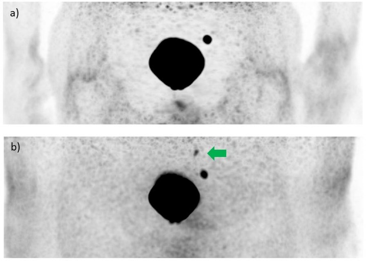
Fig 2. PET images of a patient (age: 66 years, serum PSA: 2.3 ng/ml) with recurrent PCa in two iliacal lymph nodes obtained with the PET/MR hybrid imaging system at 134 min after intravenous injection of 68Ga-HBED-CC-PSMA (146 MBq).
a) Scatter und attenuation corrected PET images with intense ‘halo artifact’ showing only one iliacal lymph node. b) Non-scatter and attenuation corrected PET image showing both lymph nodes that were seen on PET/CT as well.

