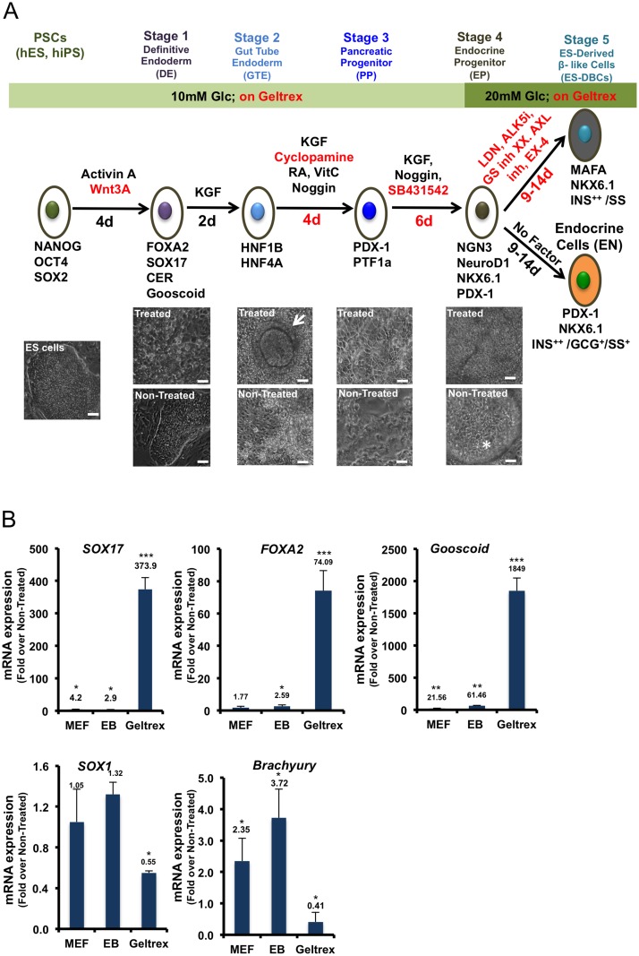
Fig 1. Short protocol outline.
(A) Schematic overview of the 25 to 30-day protocol to generate human H1 ES-derived beta-like cells (DBCs). Below, images of the differentiated H1 cells and the control cells (Non-Treated ES cell) at each stage are shown. The arrow symbol identifies tube-like structure in the differentiated cells in the stage 2. The star symbol identifies detached dead cells as spheres in the Non-Treated cells in stage 4. Scale bar = 100μm for all cell images. The red font indicates modifications to molecules or timing in comparison to the protocol described by Rezania et al [9]. (B) Expression analyses of SOX17, FOXA2 and Gooscoid as Definitive Endoderm (DE), Sox1 as ectoderm, and Brachyury as mesoderm-specific markers in the H1 ES cells differentiated on MEF, Mouse Embryonic Fibroblast; as EB (Embryoid Bodies) or on Geltrex, analyzed by quantitative RT-PCR. (* p< 0.05, **p< 0.01, p***<0.001, significant differences between the treated and control cells in each condition, unpaired two-tailed t-test, n = 3).

