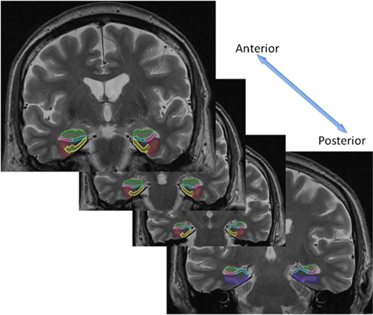
Figure 1.
Hippocampal subregions were manually drawn on the high-resolution T2 image. Shown above, moving from anterior to posterior, the subregions included the CA1 (pink), CA2, CA3 and dentate gyrus (green), the subiculum (blue), the entorhinal cortex (yellow), the perirhinal cortex (red), and the parahippocampal gyrus (purple).

