
Figure 3.
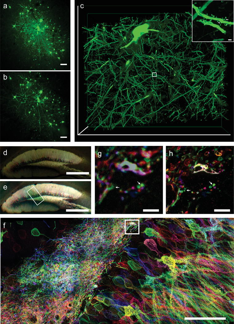
proExM of mammalian brain circuitry. (a) Wide-field image of GFP fluorescence in virally injected rhesus macaque cortex. (b) Post-expansion wide-field fluorescence image of (a). (c) Volume rendering of confocal microscopy images of subregion of (b). Inset shows a zoom-in of boxed region in (c) showing dendritic spines. (d) Low magnification widefield fluorescence imaging showing immunostained mouse hippocampus expressing virally delivered Brainbow3.0. (e) Post-expansion wide-field image of sample from (e). (f) MIP high resolution confocal microscopy image following expansion of membrane labeled Brainbow3.0 neurons from boxed region in (e). (g) Pre-expansion confocal image showing one optical section of boxed region in (f). (h) Post-expansion image of (g). Scale bars: (a) 100 μm, (b) 100 μm (physical size post-expansion, 413 μm); (c) 300 μm × 254 μm × 25 μm, (c) (i) 1 μm, (d) 500 μm, (e) 500 μm (1980 μm); (f) 5 μm, (g) 5 μm (19.8 μm); (h) 50 μm (198 μm).