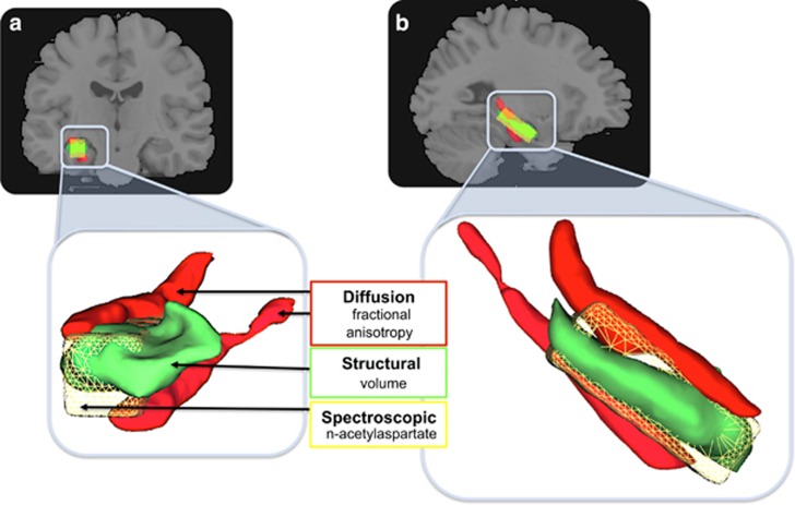
Figure 1.
Multimodal assay of the medial temporal grey matter and white matter, and biochemistry. (a) Coronal view; (b) sagittal view. Concurrent (within subjects) assay of fractional anisotropy from the fimbria and cingulum–hippocampus white-matter fibres (red); anatomical grey-matter volume of the hippocampus proper (green); and N-acetylaspartate from a voxel positioned over the hippocampus (yellow). Lower panels show a close-up rendered image to illustrate the relative positions and associations of the medial temporal regions assayed.

